There are indications that myofascial pain contributes to different types of lower urinary tract symptoms, dyspareunia and chronic pelvic pain. Pelvic physiotherapy treating myofascial pain might be a treatment option that can be considered. However, physicians are often not or not enough aware of this. Or they don’t know how to examine the pelvic floor muscles exactly on myofascial pain. Therefor it is necessary to have an evidence-based physical screening examination for myofascial pain. The objective of the research that I am going to discuss today is the development of a simple and reproducible screening examination for the levator ani and obturator internus muscle in women.
Materials and methods
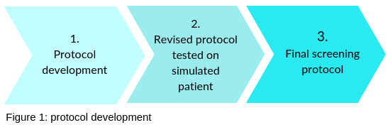
Protocol development of the screening examination was initially done by Female Pelvic Medicine and Reconstructive Surgeons (FPMRS) and Women’s health physical therapists (1). This protocol was based on items found in a systematic review (ref) and on clinical experience. This protocol was reviewed by other FPMRS physicians, adapted and tested on one simulated patient (2). Based on the feedback of the simulated patient (SP) the protocol was revised and finalized (3) (figure 1).
Standardizing the pressure applied during the investigation is not easy. However, in this research the first 16 patients were investigated with the use of a pressure sensor. An interim analysis showed that the pressure used was consistent among examiners. Therefor the pressure sensor was not used in further evaluations. Another way pressure was standardized was by palpation of the mid thigh (rectus femoris muscle). When I read this in my mind I made the analogy of the finger test for checking the doneness of a steak.
Not all professionals are familiar with an internal investigation. Therefor it was also researched if external sites can indicate myofascial pain internally. External sites were: left and right sacroiliac and anterior superior iliac spine (insertion of iliacus muscle), and insertion of the rectus abdominis muscle (pubic bone) were palpated. Every pain point scores 1 point. So the minimum score was 0 and maximum was 5. This was compared with palpation of the levator ani and obturator internus. Pain on internal palpation was based on the numeric rating scale with a 0 no pain and 10 extreme pain. These numbers were dichotomised. A score <4 was a no and ≥4 a yes. This threshold is used because it is used in the research clinic were the research took place.
New patients of the Women’s Center for Bladder and Pelvic Health at Washington University in St.Louis (USA) were asked to participate.
Inclusion criteria:
- ≥ 18 years
- Willing to participate with the research protocol
Exclusion criteria:
- Can’t rest comfortably in the research chair
- A cognitive impairment
The examinations were performed by 4 examiners in pairs of 2. They were blinded for each other’s results. (A full screening examination protocol can be found in the original article).
Internal investigation
The internal investigation of the muscles is performed with the index finger of the dominant hand. The examiners palpates counter clock-wise. Palpation starts in the muscle belly and continues in the direction of the muscle fibers, so in the length.
- Right obturator internus
- Right levator ani
- Left obturator internus
- left levator ani
Explanation how to find and palpate the obturator internus muscle:
- Place the free hand on the lateral side of the right knee (when right obturator is tested) and ask the patient to press outside. So ask for abduction. Palpate the obturator internus between 9 and 12 o’çlock.
Results
Thirty-five patients were included and screened. The median age was 55 years (range: 29-83). All women had some kind of urinary symptoms. Approximately 25% had prolapse symptoms. No patients were included with pelvic pain.
Pain scores:
High agreement (%) at all external sites (80-86%). High correlation between examiners regarding the internal investigation.
Association between pain at external sites and pelvic floor myofascial pain scores. This research showed that by palpating the external points nearly 70% of the patients with myofascial pain were identified. So palpating these points might be an option for some clinicians.
This research is a first indication that an easy, quick screening of the internal pelvic muscles is reproducible. However, to improve the robustness of this screening examination more tests are necessary with different testers and patients. More evidence is warranted concerning the relation between myofascial pain and pelvic floor symptoms.
Personal opinion & clinical implications
- This research is only for women, so there is need of a study to see if this protocol can be used for men as well.
- We can discuss this screening protocol with our network of physicians and show how this can be easily implemented.
- The external screening can also be used by colleague physiotherapists if they suspect internal myofascial pain points.
Conclusion:
This study is a first and interesting step in the development of an easy protocol which can improve a multidisciplinary approach in treating patients with lower urinary tract dysfunctions and different pelvic floor related problems. The protocol needs more testing but in the meantime it can be used to identify myofascial pain. Patients with myofascial pain can be referred to pelvic physiotherapists for a full evaluation of the pelvic muscles.
Reference:
Meister MR, Sutcliffe S, Ghetti C, et al. Development of a standardized, reproducible screening examination for assessment of pelvic floor myofascial pain. Am J Obstet Gynecol 2019; 220:255.e1-9.
Meister MR, Shivakumar N, Sutcliffe S, et al. Physical examination techniques for the assessment of pelvic floor myofascial pain: a systematic review. Am J Obstet Gynecol 2018;219:497.e1-13


Recent Comments